Home

sconosciuto greca prova gram microscope disinfettante inafferrabile Becks

Gram Stain Procedure in Research and Labs

Eisco Prepared Microscope Slide - Escherichia Coli Smear, Gram Stain  Microbiology | Fisher Scientific
Eisco Prepared Microscope Slide - Escherichia Coli Smear, Gram Stain Microbiology | Fisher Scientific
Welcome to Microbugz - Gram Stain

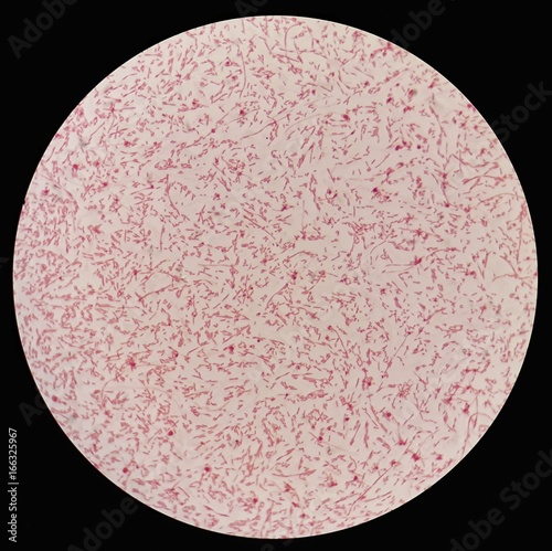
Smear of gram negative bacilli bacteria under 100X light microscope  (Selective focus). Stock Photo | Adobe Stock
Smear of gram negative bacilli bacteria under 100X light microscope (Selective focus). Stock Photo | Adobe Stock
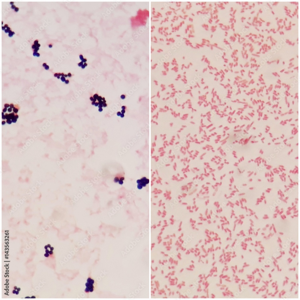
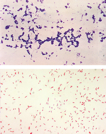
Smear of gram positive bacteria on the left and gram negative bacteria on  the right, under 100X light microscope. Stock Photo | Adobe Stock
Smear of gram positive bacteria on the left and gram negative bacteria on the right, under 100X light microscope. Stock Photo | Adobe Stock

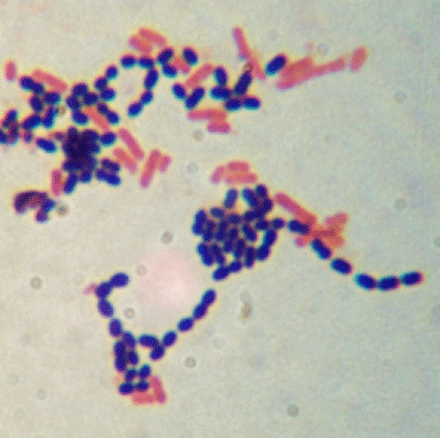
Mixed Gram-Positive and Gram-Negative Bacillus, w.m. Gram Stain Microscope  Slide | Carolina.com
Mixed Gram-Positive and Gram-Negative Bacillus, w.m. Gram Stain Microscope Slide | Carolina.com

Gram Stain Kit
Gram Stain Kit

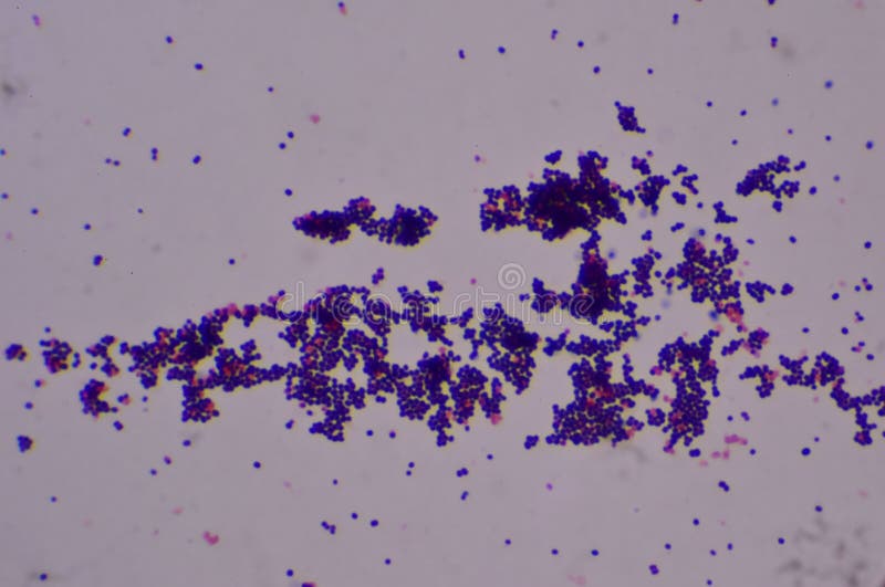
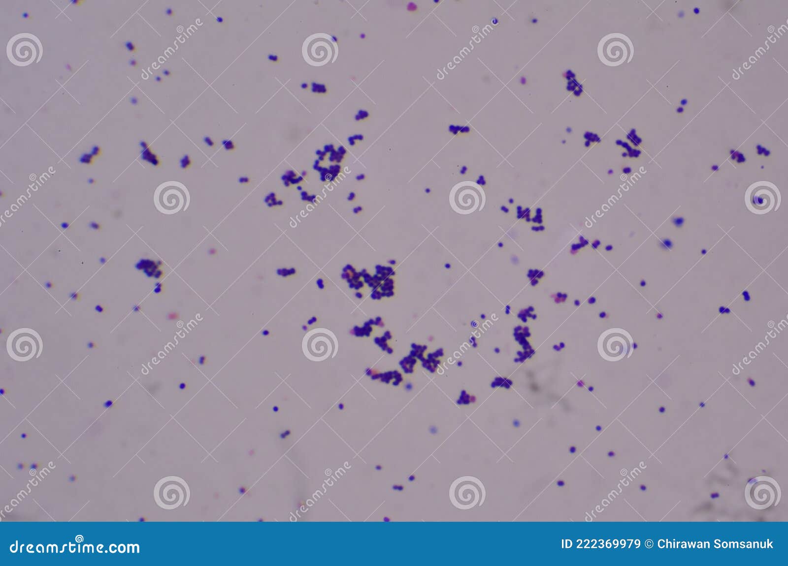
Gram Positive Cocci Finding with Microscope Stock Photo - Image of beauty,  biology: 222369974
Gram Positive Cocci Finding with Microscope Stock Photo - Image of beauty, biology: 222369974

Gram-Negative Spirillum, w.m. Gram Stain Microscope Slide: Science Lab  Microbiology Supplies: Amazon.com: Industrial & Scientific
Gram-Negative Spirillum, w.m. Gram Stain Microscope Slide: Science Lab Microbiology Supplies: Amazon.com: Industrial & Scientific
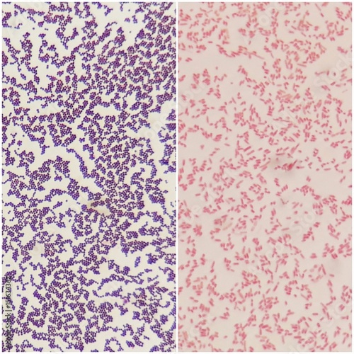
Gram Positive vs Gram Negative | Technology Networks

The Gram stain method - Labster Theory

File:Pseudomonas fluorescens Gram Stain on Microscope Slide.jpg - Wikimedia  Commons
File:Pseudomonas fluorescens Gram Stain on Microscope Slide.jpg - Wikimedia Commons

Smear of gram positive bacteria on the left and gram negative bacteria on  the right, under 100X light microscope. Stock Photo | Adobe Stock
Smear of gram positive bacteria on the left and gram negative bacteria on the right, under 100X light microscope. Stock Photo | Adobe Stock

Coccus Smear, Gram-Positive, Prepared Microscope Slide - 75x25mm - Eisco  Labs | eBay
Coccus Smear, Gram-Positive, Prepared Microscope Slide - 75x25mm - Eisco Labs | eBay

Gram Negative Rods/ Bacilli of Klebsiella pneumoniae under the Microscope -  YouTube
Gram Negative Rods/ Bacilli of Klebsiella pneumoniae under the Microscope - YouTube

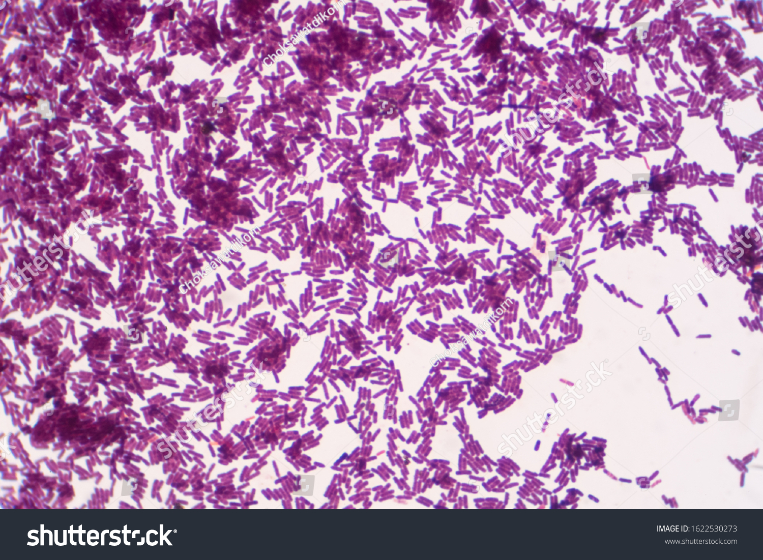
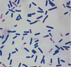
Bacillus Gram Positive Stain Under Microscope Stock Photo 1622530273 |  Shutterstock
Bacillus Gram Positive Stain Under Microscope Stock Photo 1622530273 | Shutterstock

Gram positive and Gram negative bacterial slides under the Microscope -  YouTube
Gram positive and Gram negative bacterial slides under the Microscope - YouTube

Gram Positive Cocci Finding with Microscope Stock Image - Image of  clinical, infection: 222369979
Gram Positive Cocci Finding with Microscope Stock Image - Image of clinical, infection: 222369979

Bacillus Gram Positive Stain Under The Microscope View Bacillus Is  Rodshaped Bacteria Stock Photo - Download Image Now - iStock
Bacillus Gram Positive Stain Under The Microscope View Bacillus Is Rodshaped Bacteria Stock Photo - Download Image Now - iStock

Gram-negative bacteria - Wikipedia

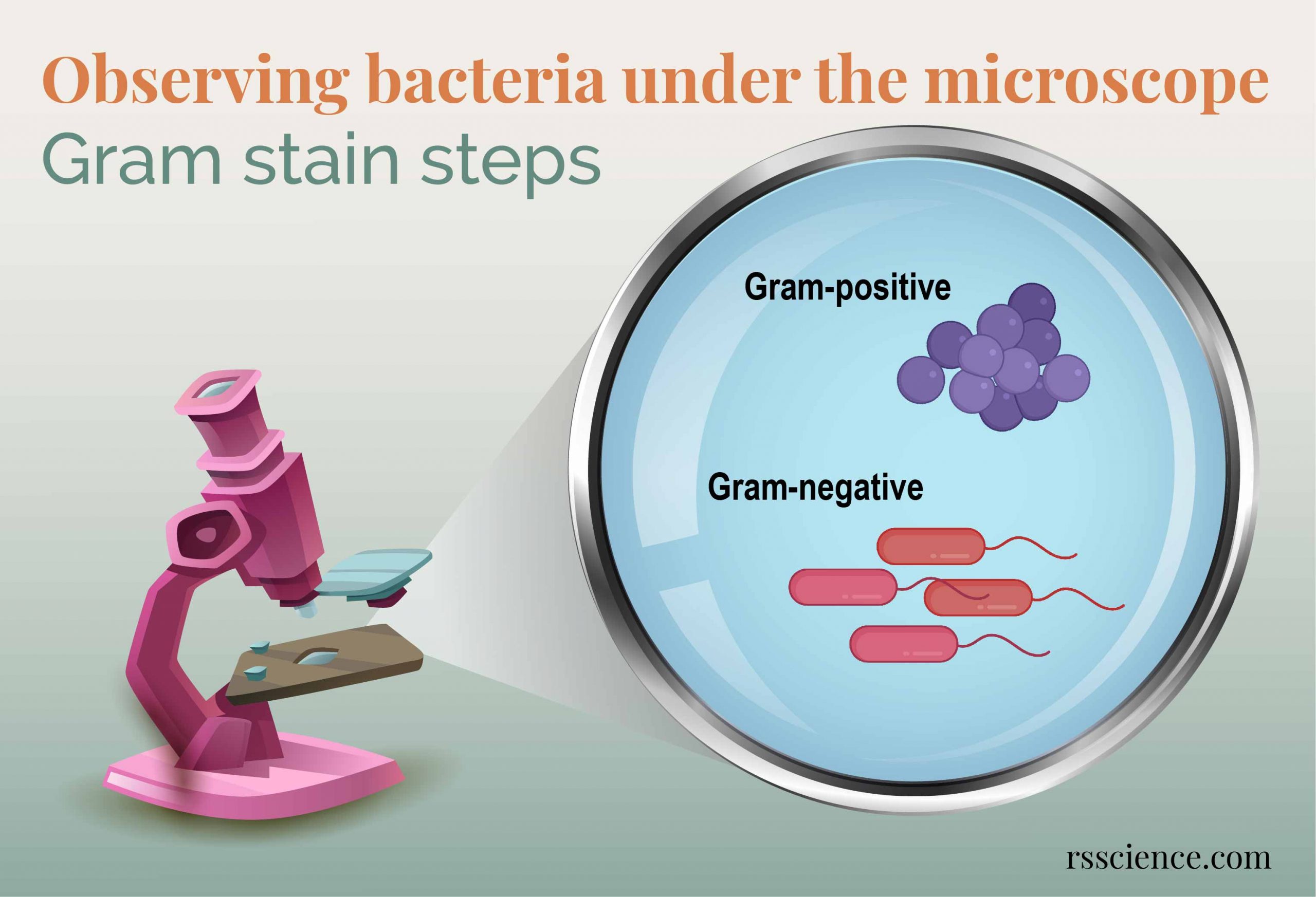
Observing Bacteria Under the Microscope - Gram Stain Steps - Rs' Science

Gram Staining Rules
Gram Staining Rules

Gram Negative Bacterial Bloodstream Infection Seen Through Microscope Stock  Photo - Download Image Now - iStock
Gram Negative Bacterial Bloodstream Infection Seen Through Microscope Stock Photo - Download Image Now - iStock

Microscope World Blog: Microscopy Gram Staining